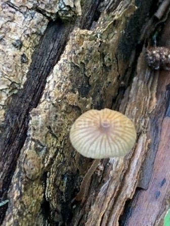
On the 18th October 2022 we noticed this tiny little mushroom growing out of a small branch of deadwood in the wetland area. We decided to attempt to identify it using the Collins Fungi Guide: Buczacki, S. Sheilds, C and Ovenden, D. Harper Collings. 2012 (Ref 13. Click Here)
We immediately realised that small brown fungi like this one are very common, with dozens of similar looking species to consider. We also quickly discovered that the fungi guide, whilst being very comprehensive, lacked a really detailed key. Relying instead on drawings for initial ballpark identification, followed by species accounts where considerably more detail is provided. As such it was important for us to gather as much detailed information as possible.
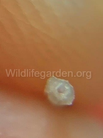
First off the overall size of the main features (the cap and stem)was taken…
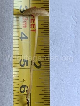

Note in the central image above the cap edge is very smooth and rounded (not wavy or dented).
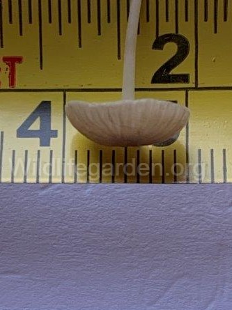
The the overall cap shape in profile is another important feature, in this case hemispherical.
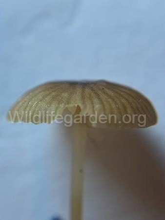
Gill attachment (how/if the gills attach to the stem) is also a key identification feature. In this case some gills don’t attach to the stem at all, something called ‘distant’. Whilst others seem to climb the stem as they attach, this is known as ‘decurrent’. Also note how crowded the gills can be as this can also be diagnostic. We considered these gills to not be very crowded based on diagrams/terms provided in the guide. The term for this level of crowding we believe is once again ‘distant’.
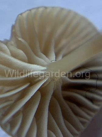
The stem was also recorded as hollow near the base of the cap. The stem lower down was to thin/soft to determine if it was solid or hollow (it crushed every time we attempted to make a clean cut).
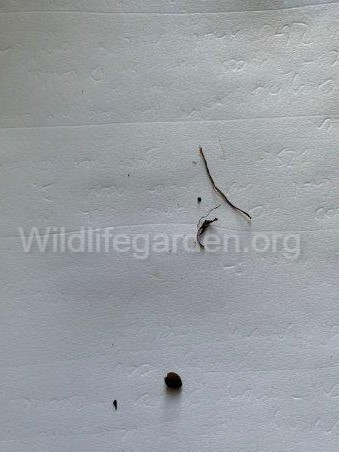
One of the key identification features is a ‘spore print’. This is obtained by carefully removing the cap from the stem. Standing the cap (gills down) on a white piece of card. Then covering with a jar to prevent drafts shifting either the cap or the spores. This allows the spores to fall from the gills leaving a pattern on the card, but crucially allowing the colour of the spores to be determined (a key identification feature). Unfortunately when we attempted this and went back 24hrs later, the cap had shrivelled to a tiny brown lump (bottom centre) with no spore pattern or anything really identifiable as a spore.
With the above information we went back to the Fungi Guide but immediately realised that without the spore colour the pictorial key was of little help as the majority of Agarics (the type of fungi we believed we were looking at) were divided up by the spore colour, with each colour representing dozens/perhaps hundreds of different species. As such we were forced to flick through the species accounts checking against each genus until after considerable time we concluded that the genus Galerina was the most likely candidate.
Even then finding a species that matched our measurements/recordings above proved difficult and as such we are recording this as simply Galerina spp. If we had to have a stab at the species, we would likely say Galerina nana, but we feel this would be pushing our luck a little.
Overall an interesting process to go through, but ultimately disappointing. We may invest in a photographic guide as well to help compliment the one we used here.
DC: 21/10/2022
